Sigmavision Advanced
Software
The interdisciplinarily-developed MR thermometry software SIGMAVISION ADVANCED is convincing due to its intelligent structure. It thus contributes to greater precision and a higher treatment quality.
Our software at a glance
- Non-invasive thermometry
- 3D temperature monitoring
- Greatest possible monitoring of the treatment
- Ergonomic, intuitive operating control with comprehensive, tried-and-tested plausibility checks
- The functional structure results from long-standing, interdisciplinary research findings and user surveys
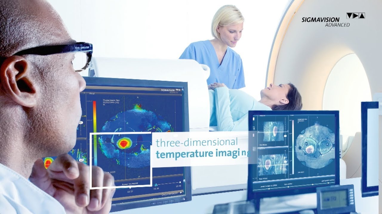
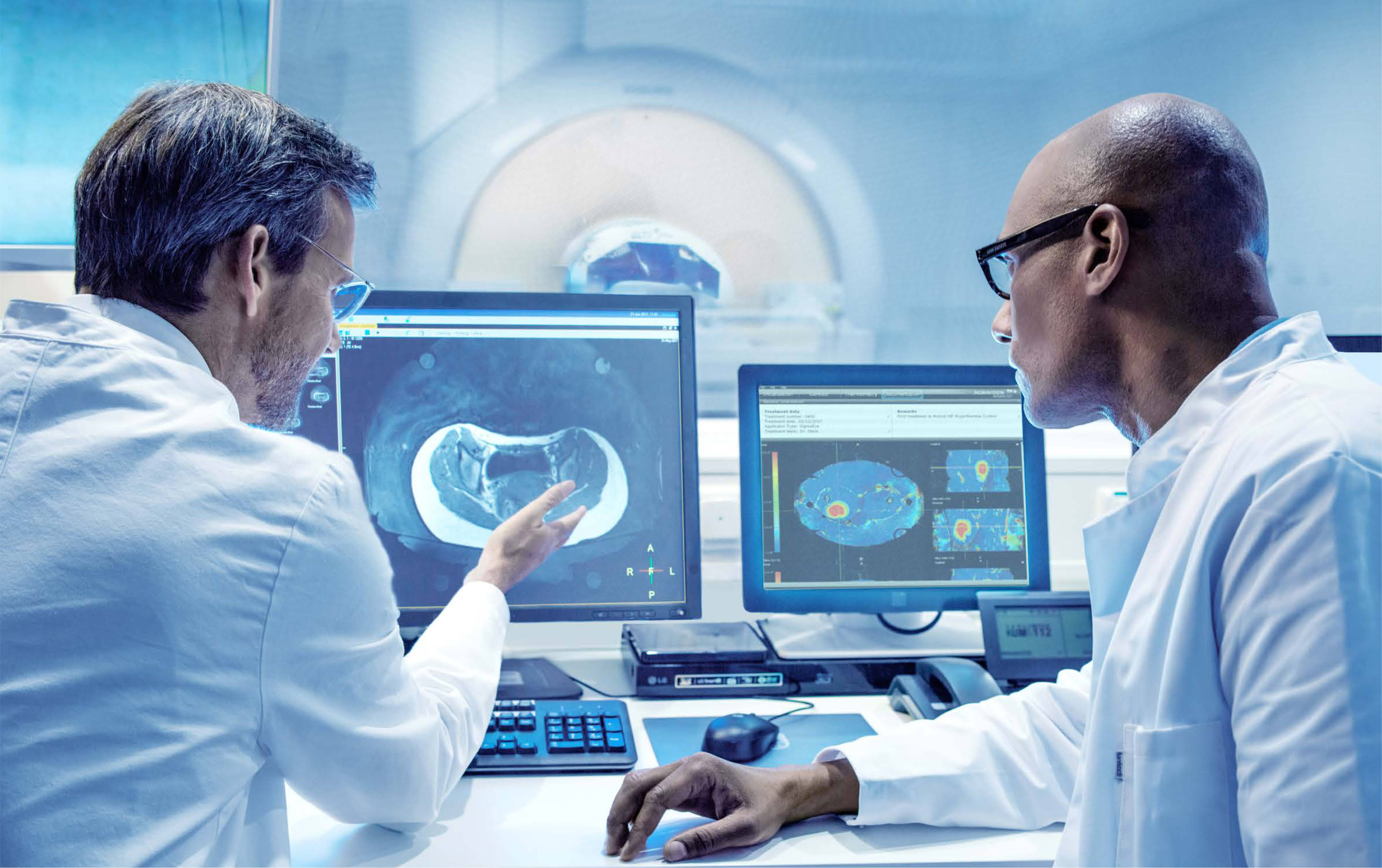
- MRI SIGMAVISION ADVANCED creates a visual image of the hybrid hyperthermia: the combination of hyperthermia and the MRI system provides a non-invasive, three-dimensional temperature imaging during the course of treatment. By monitoring the temperature change, users can react to fault heating or hot spots, which can reduce side effects.
- SIGMAVISION ADVANCED is an optimized thermometry software enabling the user to measure, monitor, and document the temperature changes
- The thermometry process is based on an algorithm developed in close cooperation with the Klinik für Radioonkologie und Strahlentherapie, Charité Universitätsmedizin in Berlin
Details
Automatic Recording
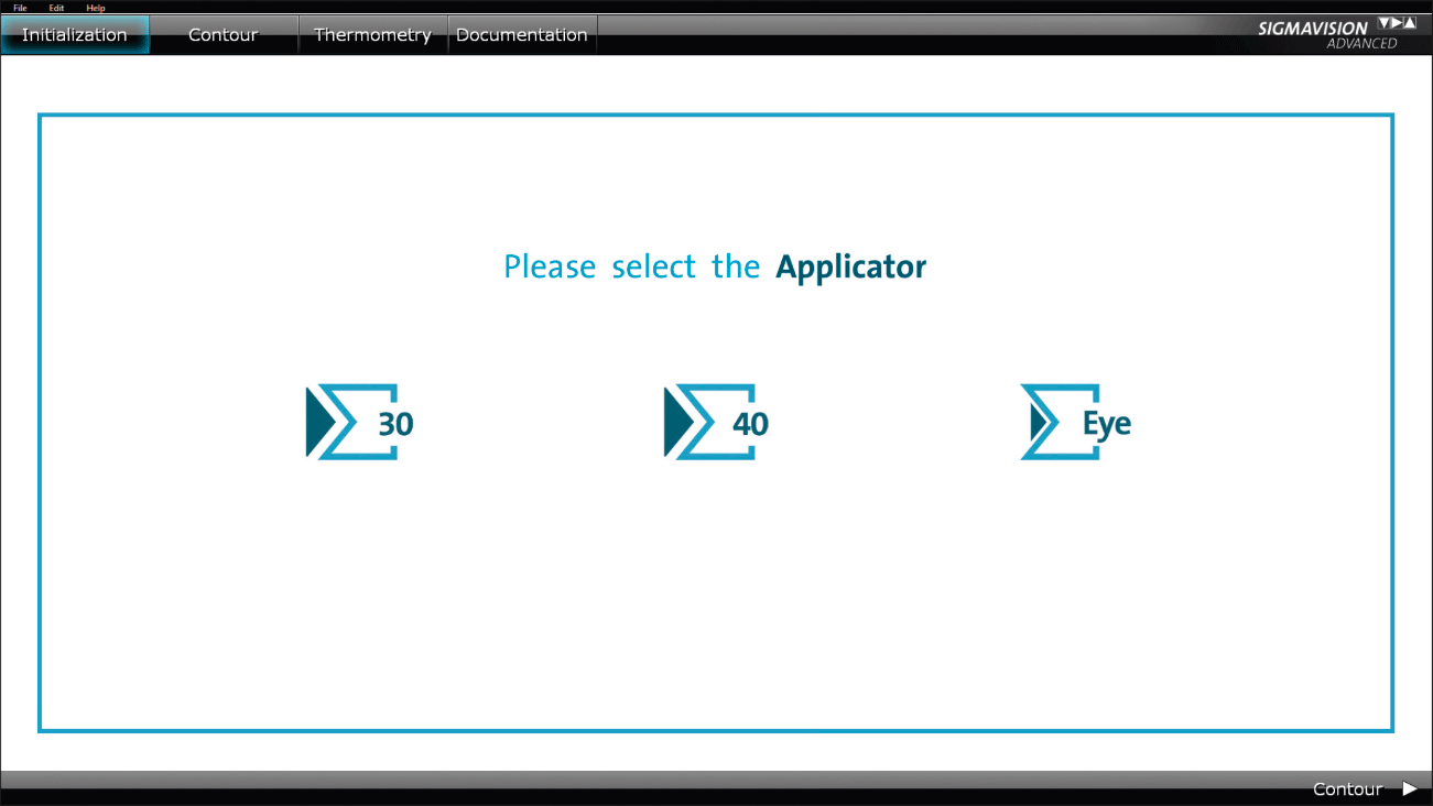
In every hybrid hyperthermia, an MRI protocol developed specially for MR thermometry is used for imaging.
- An easily understandable user interface supports the user during the import of the patient data.
- The DICOM data from the MR system can be sent to SIGMAVISION ADVANCED with a “click”. SIGMAVISION ADVANCED processes this automatically.
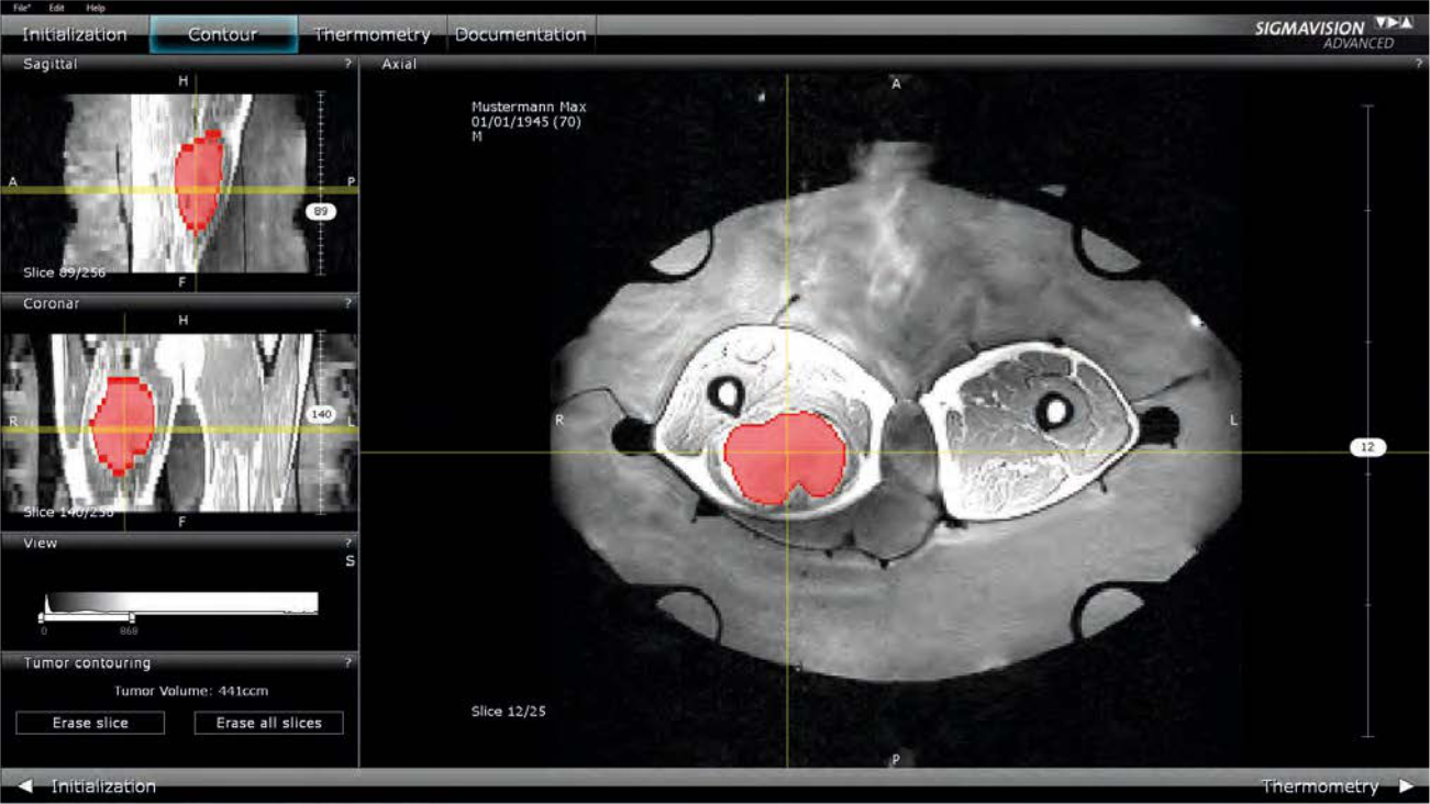
Efficient Tumor Contouring in 3D
The tumor can be contoured in SIGMAVISION ADVANCED for evaluation.
- Thanks to the three-dimensional MRI images, SIGMAVISION ADVANCED enables 3D editing of the contours.
- The axial, sagittal, and coronal slices of the MRI images are displayed on the screen.
- This is followed by an automatic correction of the magnetic field drift of the MRI system.
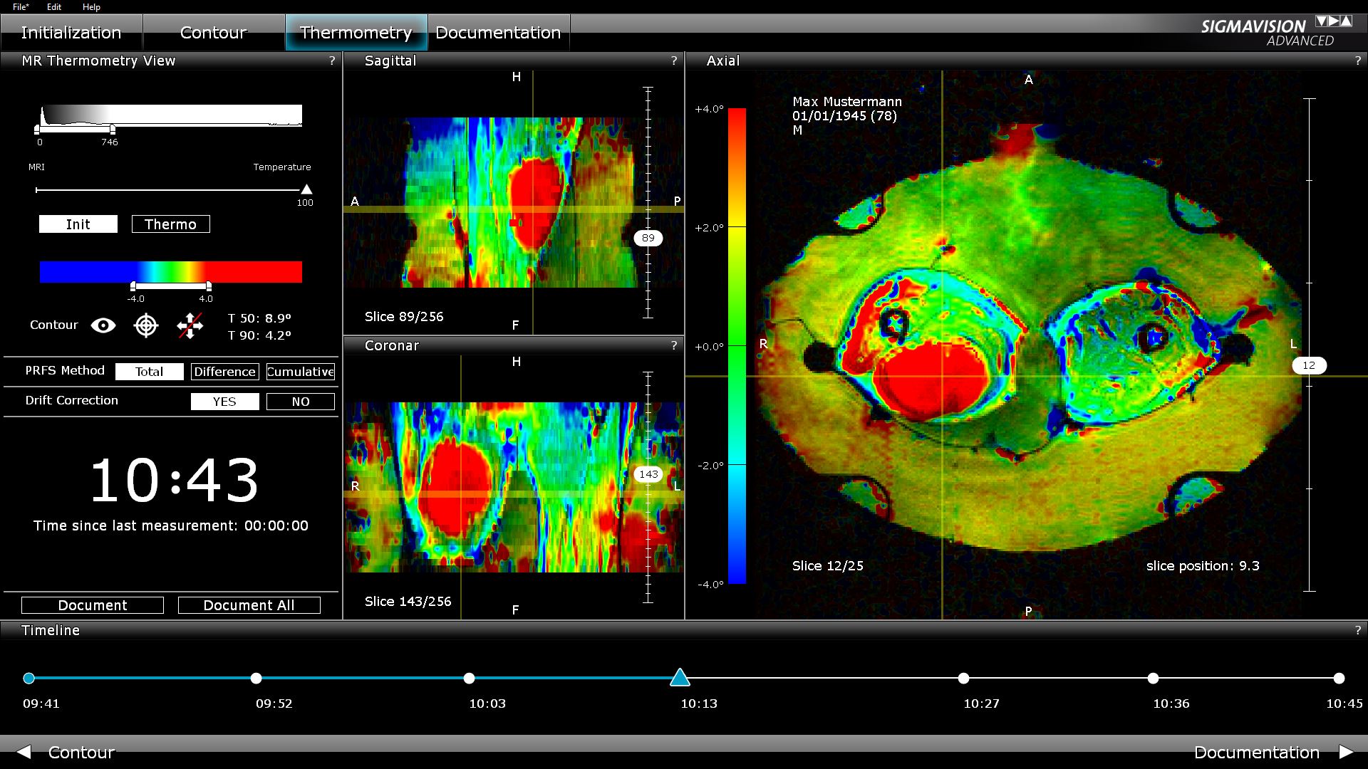
Non-invasive Temperature Monitoring during the treatment
This menu point allows for the calculation and visualization of the axial thermal images. Following each new MRI measurement, the user is supplied with the calculated, color-coded 3D temperature images.
During treatment, the MR-image-guided hyperthermia records the MR volume at regular intervals – e g every 5 minutes – and color-coded regions represent the calculated thermal image on the resulting axial slices. The temporal progression of the temperature changes in the tumor and muscle tissue can be reconstructed via the timeline. The user can react to unexpected and undesired heating (hot spots) during the treatment.
SIGMAVISION ADVANCED represents a highly professional tool for the recording of heat changes in the patient during the treatment.
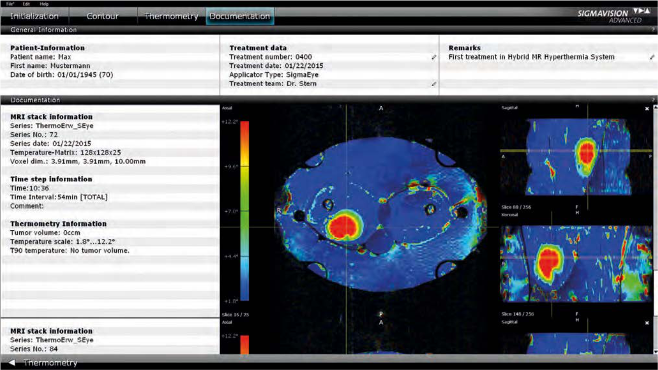
Every Detail Saved
SIGMAVISION ADVANCED collates all MR thermometry data and settings, from the patient data to the contouring of the tumor, right up to the applicator data and the graphical evaluation of the temperature measurement. These parameters can be saved and printed out properly formatted.