Currently not available for sale in Germany and Austria.

Examples
use cases

Point-of-Care Imaging in Neurocritical Care Settings

Ultimately sequence choice and plane selection decisions should be made with the clinical and radiology teams in consultation together, and should be selected based on the clinical question to be answered. Below are example use cases and the sequences physicians have found useful in their examinations for producing images that provided information relevant to the clinical questions listed. This information is meant to be a reference only.

Studies show that clinicians can use the Swoop system at the point of care for assessing:
change in patient symptoms with an unknown cause1,3,5,6, follow-up scans for clinically suspected or known strokes greater than 5mm2, change in ventricular size with or without intervention6,7, change in an intraparenchymal hematoma2,8, change in extra-axial collection7, change in the imaging appearance of infarct1, to follow or confirm stability4, mass effect and potential for midline shift9, and change post‐thrombectomy10.

1. Post-operative Trauma Follow-up Assessment

:
Run based on which sequence(s) the pathology has been previously best seen.

:
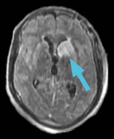
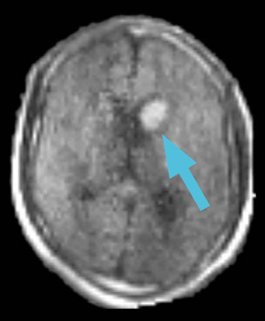
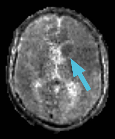
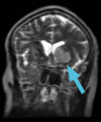
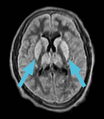
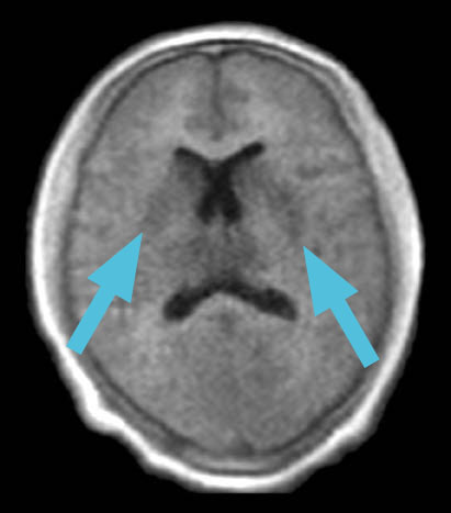
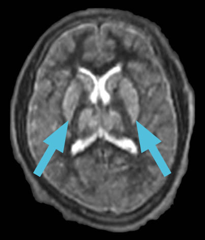
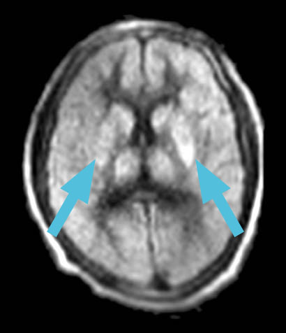
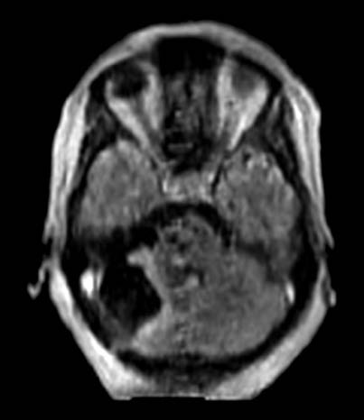
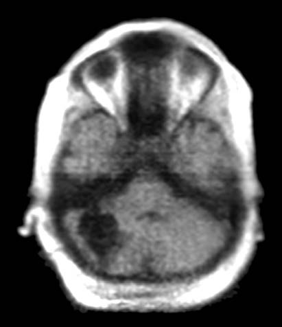
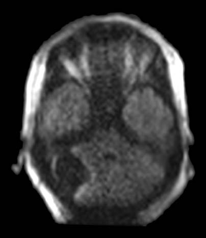
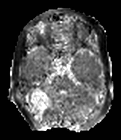
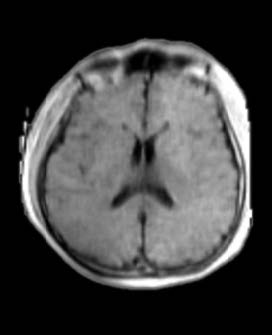
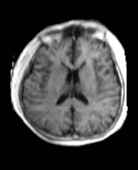
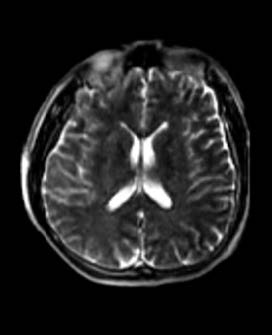
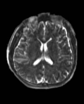
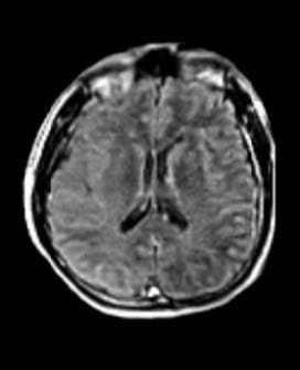
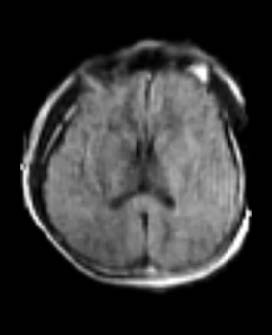
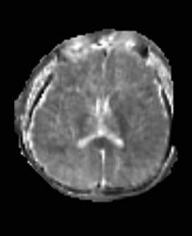
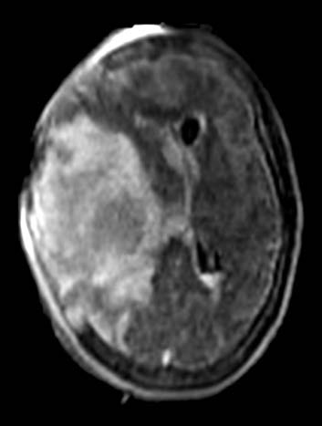
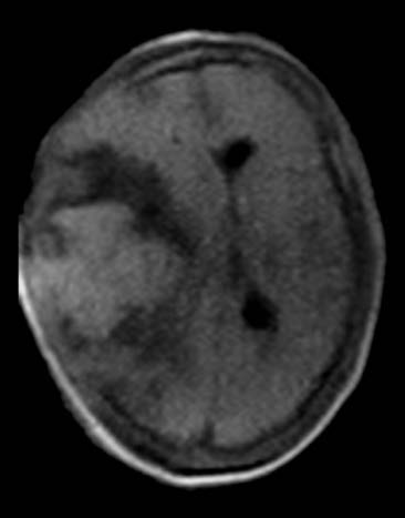
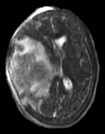
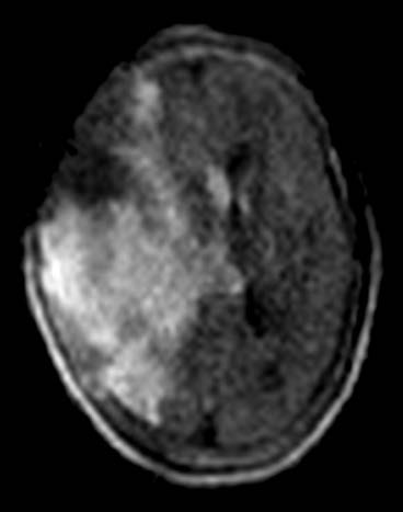
A 35-year-old female found unresponsive was brought into the emergency room. An emergent head CT revealed a massive traumatic intraparenchymal hemorrhage. Clinicians immediately took the patient to the operating room for decompression.

:
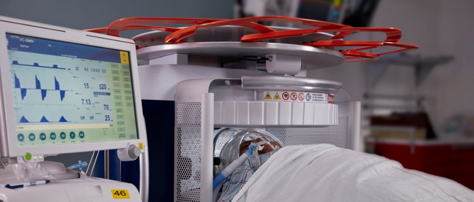
Post-operatively, physicians used the Swoop system at the patient’s bedside. The images assisted them in assessing the extent of mass effect, midline shift, and tissue viability for prognostication.

FLAIR
T1 (Standard)
Fast T2
DWI

The Swoop system is not intended to apply color overlays to images. Colors are added for clarity and are not reflective of the original images.

2. Post-op Infarct

:
FLAIR, DWI with ADC, T1 (Standard), Fast T2

:
A 56-year-old male with a history of prior transsphenoidal pituitary resection recently underwent a pterional approach for additional resection. On post-op day one, the patient experienced a seizure and coded, presenting with new neurological signs, including right-sided weakness and a non-responsive pupil.

:
Swoop system images assisted the physicians in promptly diagnosing this unstable, immediate post-operative patient.

FLAIR
DWI
ADC
T1 (Standard)
T2

The Swoop system is not intended to apply color overlays to images. Colors are added for clarity and are not reflective of the original images.

3. Post-cardiac Arrest Anoxia

:
Run based on which sequence(s) the pathology has been previously best seen.

:
A 31-year-old man, post-drug overdose, presented in the ED and experienced cardiac arrest. Physicians administered naloxone for opioid reversal. Critically ill and unresponsive, having undergone prolonged resuscitation, the patient was transferred to the ICU.

:
Swoop system images, taken after five days, assisted the physicians in identifying cerebral anoxia and sub-acute infarcts in the deep gray matter of the dorsal midbrain, bilateral thalami, and basal ganglia, guiding the decision to withdraw life support.

FLAIR
T1 (Standard)
FAST T2
DWI

The Swoop system is not intended to apply color overlays to images. Colors are added for clarity and are not reflective of the original images.

4. Post-operative ICU Exam

:
Run based on which sequence(s) the pathology has been previously best seen.

:
A 39-year-old female with a history of recurrent metastatic non-small cell lung carcinoma. She now presents with a growing right-sided posterior fossa mass. Physicians used the Swoop system in the ICU twelve hours after surgery to assess her condition.

:
Swoop system images showed a total resection with no evidence of hemorrhage, significant edema, mass effect, or obstructive hydrocephalus. Following the exam, physicians transferred the patient out of the ICU on postoperative day one, saving time and cost.

FLAIR
T1 (Standard)
DWI
ADC

The Swoop system is not intended to apply color overlays to images. Colors are added for clarity and are not reflective of the original images.

Swoop System Sequences

The Swoop system offers T1, T2, fluid-attenuated inversion recovery (FLAIR), and diffusion-weighted imaging (DWI) with apparent diffusion coefficient (ADC) map sequences (with two T1 and T2 variations). The T1, T2, and FLAIR sequences are available in the axial, coronal, and sagittal planes.

T1 (Standard)

1.6 × 1.6 × 5 mm

T1 (Gray/White)

1.6 × 1.6 × 5 mm

T2

1.5 × 1.5 × 5 mm

Fast T2

1.6 × 1.6 × 5 mm

FLAIR

1.7 × 1.7 × 5 mm

DWI

2.4 × 2.4 × 6 mm

DWI (with ADC Map)

2.4 × 2.4 × 6 mm

Endnotes

  1. Kuoy E, Glavis-Bloom J, Hovis G, et al. Point-of-Care Brain MRI: Preliminary Results from a Single-Center Retrospective Study. Radiology. 2022;305(3):666-671. doi:10.1148/radiol.211721

  2. Mazurek MH, Cahn BA, Yuen MM, et al. Portable, bedside, low-field magnetic resonance imaging for evaluation of intracerebral hemorrhage. Nat Commun. 2021;12(1):5119. Published 2021 Aug 25. doi:10.1038/s41467-021-25441-6

  3. Turpin J, Unadkat P, Thomas J, et al. Portable Magnetic Resonance Imaging for ICU Patients. Crit Care Explor. 2020;2(12):e0306. Published 2020 Dec 21. doi:10.1097/CCE.0000000000000306

  4. Yuen MM, Prabhat AM, Mazurek MH, et al. Portable, low-field magnetic resonance imaging enables highly accessible and dynamic bedside evaluation of ischemic stroke. Sci Adv. 2022;8(16):eabm3952. doi:10.1126/sciadv.abm3952

  5. Sheth KN, Mazurek MH, Yuen MM, et al. Assessment of Brain Injury Using Portable, Low-Field Magnetic Resonance Imaging at the Bedside of Critically Ill Patients. JAMA Neurol. Published online September 8, 2020. doi:10.1001/jamaneurol.2020.3263

  6. Beekman R, Crawford A, Mazurek MH, et al. Bedside monitoring of hypoxic ischemic brain injury using low-field, portable brain magnetic resonance imaging after cardiac arrest. Resuscitation. 2022;176:150-158. doi:10.1016/j.resuscitation.2022.05.002

  7. Sien ME, Robinson AL, Hu HH, et al. Feasibility of and experience using a portable MRI scanner in the neonatal intensive care unit. Arch Dis Child Fetal Neonatal Ed. 2023;108(1):45-50. doi:10.1136/archdischild-2022-324200

  8. Mazurek MH, Parasuram, NR, Peng TJ et al. Detection of Intracerebral Hemorrhage Using Low-Field, Portable Magnetic Resonance Imaging in Patients With Stroke. Stroke. 2023;54:2832–2841. doi:10.1161/STROKEAHA.123.043146

  9. Sheth KN, Yuen MM, Mazurek MH, et al. Bedside detection of intracranial midline shift using portable magnetic resonance imaging. Sci Rep. 2022;12(1):67. Published 2022 Jan 7. doi:10.1038/s41598-021-03892-7

  10. Sujijantarat N, Koo AB, Jambor I, et al. Low‐Field Portable Magnetic Resonance Imaging for Post‐Thrombectomy Assessment of Ongoing Brain Injury. Stroke. 2023;3:e000921. Published 2023 Jul 24. doi:10.1161/SVIN.123.000921

This content is for general information only and is not intended to substitute for formal medical training or certification. Hyperfine, Inc. does not provide clinical training, nor does it provide or evaluate physician credentialing or train physicians in procedures or techniques. Before performing an MR scan, physicians are responsible for receiving sufficient training and proctoring to ensure that they have the skill and experience necessary to protect the health and safety of the patient. For technical information, including full cautions and warnings on using the Swoop system, please refer to the instructions for use (LBL-000339). Read all instructions carefully. Failure to properly follow instructions, notes, cautions, warnings, and danger messages associated with this equipment may lead to serious injury or complications for the patient. While clinical studies support the use of the Swoop system for portable cranial magnetic resonance imaging, individual results may vary.